


INTRODUCTION
Traumatic pseudoaneurysms, which are uncommon vascular lesions arising from traumatic injuries to the nasal and facial areas [1], pose a significant clinical challenge due to their potential to cause recurrent and life-threatening epistaxis [2]. Pseudoaneurysms develop due to direct arterial injury, leading to a hematoma within the adjacent tissues [3]. Although uncommon, traumatic sphenoid sinus pseudoaneurysms originating from the cavernous segment of the internal carotid artery require immediate recognition and proper management to avert serious complications. Understanding the underlying vascular anatomy, causes, and treatment options is essential for optimizing patient outcomes.
In this case report, we present a 55-year-old man who developed a traumatic sphenoid sinus pseudoaneurysm originating from the cavernous segment of the internal carotid artery following a traumatic incident on April 10, 2023. The patient experienced recurrent and persistent epistaxis, which necessitated emergent medical intervention. This led to the successful treatment of the pseudoaneurysm through stent-assisted coil embolization.
CASE REPORT
A 55-year-old man presented to the emergency department on May 10, 2023, with a history of recurrent epistaxis following an incident involving a wire on April 10, 2023 (i.e., 31 days prior). The patient had a history of acute cerebral infarction, which occurred after the trauma, but no other significant past medical history was reported. He described the trauma as an incident where a wire penetrated his nose. After the initial trauma, the patient had experienced repeated episodes of nasal bleeding. The traumatic event, which occurred approximately one month earlier, caused persistent discomfort and intermittent episodes of bleeding.
Upon examination, the patient’s medical history revealed no previous episodes of epistaxis or significant nasal symptoms. The patient’s vital signs were stable, indicating no signs of an acute event. The initial laboratory findings showed a hemoglobin level of 7.5 g/dL, prompting a consultation with the hematology/oncology department to evaluate the finding of anemia. Nasal endoscopy revealed active bleeding from the posterior nasal region, which raised concerns about potential underlying vascular issues. Initial attempts to control the bleeding with conservative measures, such as nasal packing, were unsuccessful.
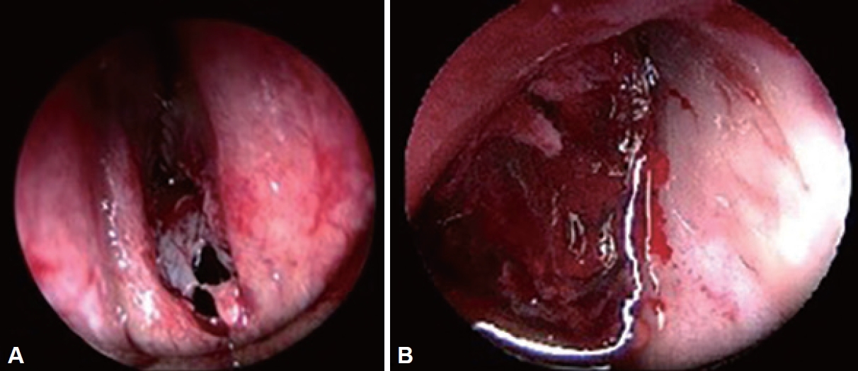
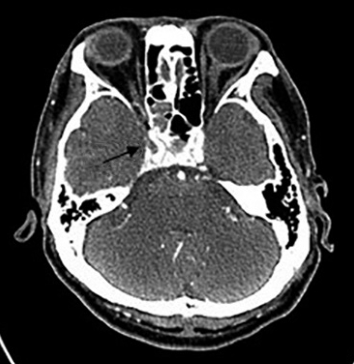
The patient’s persistent and recurrent epistaxis necessitated further management. The patient underwent an emergency operation 31 days after a traumatic event. During the operation, a pulsating soft tissue mass, suspicious for a pseudoaneurysm, was observed within the sphenoid sinus, arising from the posterior wall (Fig. 1). The operation was concluded upon identification of the pulsating mass, and a radiologic evaluation was planned. During the postoperative observation period, there was no evidence of cerebrospinal fluid rhinorrhea. Urgent computed tomography angiography (CTA) of the carotid arteries was performed to assess the vascular structures (Fig. 2). CTA revealed a pseudoaneurysm originating from the cavernous segment of the internal carotid artery near the trauma site (Supplementary Video 1 in the online-only Data Supplement). This pseudoaneurysm was considered the likely cause of the recurrent bleeding episodes. Given the high risk of complications associated with the pseudoaneurysm, a multidisciplinary team—including otolaryngologists and neurosurgeons—convened to discuss the optimal treatment strategy. Taking into account the patient’s clinical presentation, the team decided to perform stent-assisted coil embolization to address the pseudoaneurysm and prevent further bleeding.
Thirty-two days after the traumatic event, the patient underwent stent-assisted coil embolization of a pseudoaneurysm originating from the cavernous segment of the internal carotid artery (Fig. 3). This procedure was carried out under local anesthesia with image guidance. A microcatheter was guided to the pseudoaneurysm, and a stent was meticulously placed to facilitate the coil embolization. The intervention was successful in securing hemostasis and precluding further active bleeding.
After the procedure, the patient was closely monitored in the intensive care unit. He experienced no immediate complications and reported a resolution of the recurrent epistaxis. Follow-up imaging confirmed the successful embolization of the pseudoaneurysm, with no evidence of residual or recurrent bleeding.
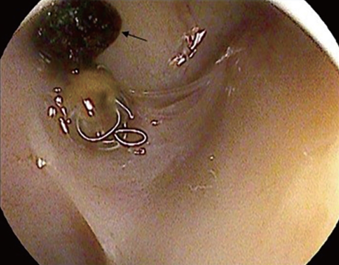
In the weeks following treatment, the patient’s condition stabilized, and there was no recurrence of epistaxis. Consequently, he was discharged from the hospital. Six months later, the patient visited an outpatient clinic, reporting no complications or recurrence of symptoms. Nasal endoscopy revealed a coil within the sphenoid sinus (Fig. 4). Further evaluation determined that the cause of the patient’s anemia was recurrent nasal bleeding. His hemoglobin levels returned to normal, measuring 13.4 g/dL.
DISCUSSION
The presented case underscores the importance of timely intervention for pseudoaneurysms that develop after traumatic events [1]. Pseudoaneurysms arise from direct arterial injury, leading to a hematoma within the adjacent tissues [3]. The patient’s initial trauma, caused by an incident involving a wire, led to ongoing discomfort and repeated episodes of epistaxis [2]. Despite initial conservative management strategies, such as nasal packing, the patient’s symptoms persisted, necessitating further evaluation.
Radiological imaging, including CTA, played a crucial role in confirming the diagnosis of a traumatic sphenoid sinus pseudoaneurysm originating from the cavernous segment of the internal carotid artery [4,5]. The occurrence of a pseudoaneurysm in the sphenoid sinus from the cavernous segment of the internal carotid artery is very rare [6,7]. The challenge in identifying the precise location of the bleeding focus stems from the fact that this segment of the internal carotid artery is situated within the cavernous sinus. However, anatomically, the cavernous segment of the internal carotid artery is located on the lateral side of the sphenoid sinus, and it is separated from the sinus by a bone barrier that is less than 0.5 mm thick [8]. The CTA findings revealed a distinct vascular lesion at the trauma site, confirming the suspicion of a pseudoaneurysm as the likely cause of the recurrent epistaxis. Prior to surgery, the patient had a history of trauma and symptoms of recurrent epistaxis, necessitating the evaluation of CTA to assess for a pseudoaneurysm or vascular injury. Angiography can serve as another gold standard for the diagnosis of a pseudoaneurysm [9]. Both computed tomography and magnetic resonance imaging can also provide valuable information regarding the potential presence of a pseudoaneurysm.
In cases with post-traumatic injuries similar to those of our patient, a pseudoaneurysm of the internal carotid artery or direct trauma to the artery itself can lead to cerebral infarction. Injury to the cavernous segment of the internal carotid artery can lead to symptoms such as diplopia, ptosis, facial pain, reduced visual acuity, and intracerebral hemorrhage. Such cases necessitate emergency intervention [10,11].
A multidisciplinary approach involving otolaryngologists, neurosurgeons, and interventional radiologists was pivotal in determining the optimal treatment strategy. In contrast to an idiopathic aneurysm, which has a true aneurysmal wall, a pseudoaneurysm lacks such a structure and cannot be packed with detachable coils [12]. Stent-assisted coil embolization was identified as the preferred method for treating the pseudoaneurysm [1,9]. This minimally invasive procedure effectively achieved hemostasis and prevented further bleeding. In our case, the patient underwent stent-assisted coil embolization for a pseudoaneurysm originating from the cavernous segment of the internal carotid artery. A microcatheter was guided to the pseudoaneurysm, and a stent was meticulously placed to facilitate the coil embolization.
The success of stent-assisted coil embolization underscores the significance of interdisciplinary collaboration and the utilization of sophisticated endovascular techniques. To evaluate the long-term efficacy of the procedure and to detect any possible recurrence, follow-up appointments were systematically conducted.
We conducted a literature review of previous cases (Table 1). Three cases [13-15] were treated through ligation of the artery, and two cases [16,17] were treated with embolization. Complications following treatment were reported in two cases [14,18], including dysphagia, right hemiparesis, and recurrent pseudoaneurysm rupture. In our case, the patient was diagnosed with cerebral infarction following trauma. Further research is needed regarding the relationship between cerebral infarction and penetrating trauma.
If diagnosis and treatment are delayed, life-threatening epistaxis, unilateral vision loss due to hemorrhage in the cavernous sinus, and subarachnoid hemorrhage can occur. A delay in diagnosis may contribute to increased morbidity and mortality in patients [19].
In conclusion, pseudoaneurysms arising from the cavernous segment of the internal carotid artery can lead to recurrent and life-threatening epistaxis [3,20]. They may also cause other complications, such as cerebral infarction, as observed in our case. Timely diagnosis and a multidisciplinary treatment approach, as demonstrated in this case, are crucial for achieving successful outcomes and preventing further complications. Patients who have experienced trauma with subsequent cerebral complications or epistaxis should undergo further evaluation for direct vascular injury or pseudoaneurysm, as in our case. Further research and case reporting are essential to improve our understanding of these rare vascular lesions and to refine the treatment strategies.

 PDF Links
 PubReader
 ePub Link
 Full text via DOI
 Download Citation
 Supplement
 Print
Print


