


INTRODUCTION
Accounting for 80% to 90% of all animal bites, dog bites are one of the most common types of trauma [1,2]. The scope of head trauma caused by a dog bite greatly varies [3,4]. In case of a massive tissue defect, it may lead to serious change in the patient’s appearance, which can be accompanied by lower quality of life and mental issues [3,5,6]. Severe traumas require reconstruction. The nose requires particularly careful and precise reconstruction due to its structural complexity and functional significance [3,7]. A wide range of reconstruction techniques have been reported for nasal tissue defect caused by a burn or a surgery to remove malignant tumor, including skin graft, local flap, and free flap [1,3-6,8-11]. Among those methods, the forehead flap can be considered as a reconstruction method for patients severely damaged by dog bites, as the technique offers a sufficient amount of tissue and poses low risk of infection [4]. Reconstruction of noses damaged by dog bites using forehead flaps has been reported in several articles [3,4,8]. In Korea, a single paper reported nasal reconstruction after a dog bite. However, in the case, a free flap was used for reconstruction [11]. Therefore, in this study, we will share the reality of dog bite treatment by sharing a case report for a patient who received a nasal construction surgery using a forehead flap.
CASE REPORT
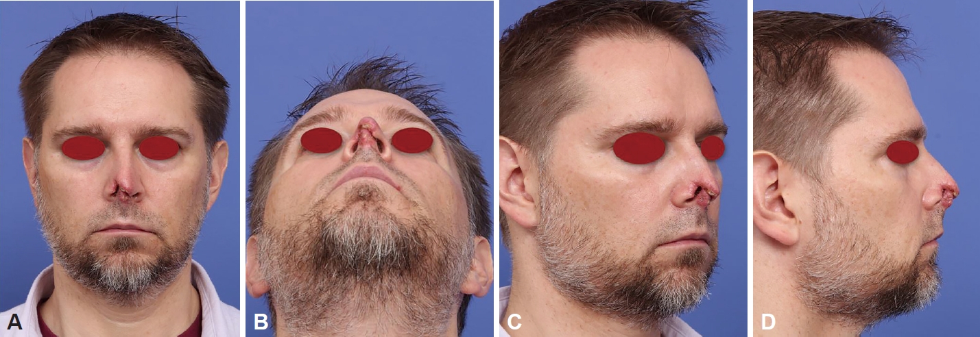
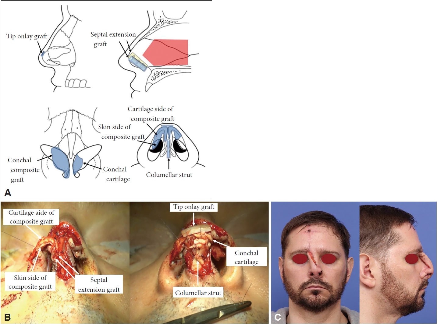
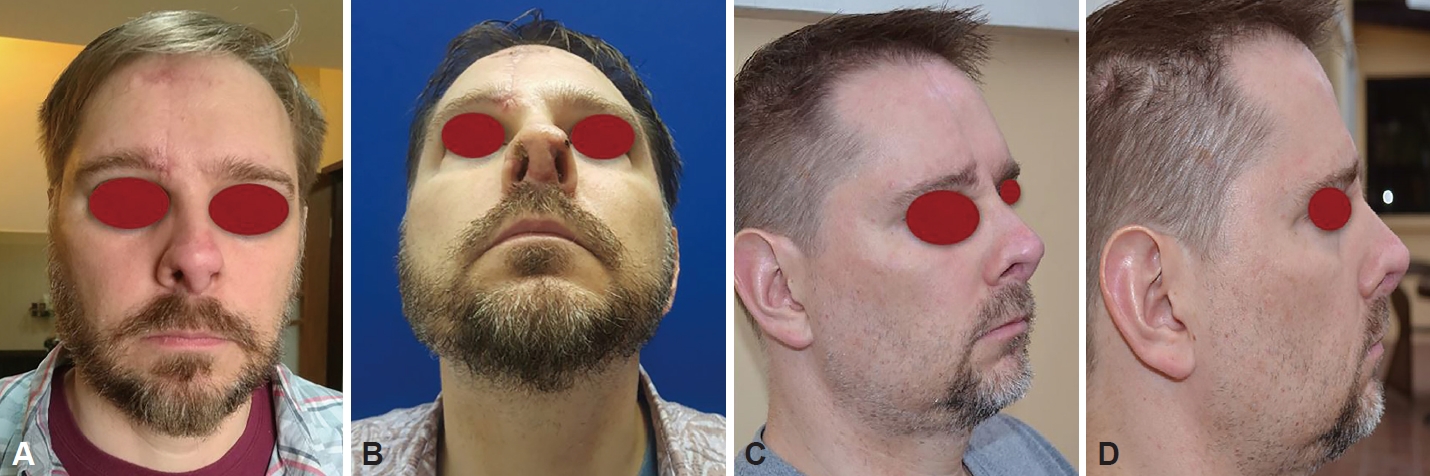
A 45-year-old United States national with non-specific findings in medical history had been bitten on the nose by a neighbor’s dog three weeks while living in the Philippines. He received conservative treatment including antibiotics at a local hospital, and then referred for a nasal construction surgery. At the time of his visit, he was identified with the loss of the upper half of his columella, defects of the skin and cartilage portion of the right lower lateral cartilage, and necrotic tissue with atrophy in the left medical crus of the lower lateral cartilage (Fig. 1). An examination with a nasal endoscopy found no additional defects in the tissues in the nasal cavity. A pre-surgery CT scan showed no specific findings other than a deviated nasal septum on the right side. The surgery was performed with general anesthesia. The skin was incised along the boundary of the non-defect area, and the dead skin on the tip of the nose was removed. To reconstruct the bone structure in the 1/3 lower section of the nose, cartilage and bone were collected from the nasal septum and a septal extension grafting was performed on the caudal end on either side. The right lower lateral cartilage and the skin inside the nose, which showed defects of skin, cartilage, and mucous membrane were reconstructed using the conchal cartilage composite graft from the right ear. The inner skin and the lower lateral cartilage were reconstructed at the same time by having the skin side of the composite graft face the inner side of the nose, and inserting and closing both ends between the patient’s lower lateral cartilage that had remained intact. By this method, the defect site of the right frontal nasal mucosa was constructed using the skin of the composite graft. The defect site of the medial crura of the left lower lateral cartilage was reconstructed using the cartilage from the left ear. Additionally, a tip only graft and a columellar strut were inserted using the ear cartilage (Fig. 2). After reconstructing the lost 1/3 bottom of the membrane and the cartilage, the right forehead flap was elevated and grafted on the defect site to reconstruct the skin and soft tissue defect sites. To obtain the forehead flap, the supratrochlear artery located 2 cm from the center line of the face was identified using Doppler ultrasound, and a 1.5 cm-thick pedicle was designed around the site. Then, after measuring the size of the defect site and the distance to the site, a flap was obtained over the prefrontal area. Then, after removing most of the frontalis muscle and fat tissue from the flap, the flap was positioned on the non-defect site and closed. The forehead flap was successfully transplanted, and a pedicle division was performed after around three weeks under local anesthesia. The patient returned to his country of residence, and recovery was confirmed by the postoperative photographs sent by the patient six months after the surgery (Fig. 3).
DISCUSSION
Dog bite is one of the most common types of animal bite. It accounts for between 26.8% and 56.5% of head and neck traumas [1,2]. Around 1% to 2% of bite patients require hospitalization [7]. The most common bite site on the head and neck is the upper lip at 37.8%, and the nose takes up around 20.6% [7]. Damages caused by bites greatly vary, from laceration to wide tissue defect. These require antibiotic treatment, and severe cases require surgical treatment. It has been reported that around 6.8% of the patients requiring surgical treatment require reconstruction surgeries beyond simple closure [7].
The nose has a complex structure, takes up a large part in a person’s appearance, and performs various roles including breathing and smelling. For this reason, it is highly difficult to reconstruct the functions and appearance of noses with tissue defects caused by burn, trauma, or malignant tumor. Unless fully reconstructed, multiple surgeries are required in some cases [3,5,8,12]. A nose with a small tissue defect site can be reconstructed using primary closure and skin grafting [3,8,13]. However, in case of larger defect sites, a sufficient amount of tissue is required, and a forehead flap or a free flap may be needed for reconstruction [1,3-6,8-11]. Forehead flap is widely used for the reconstruction of noses with tissue defect caused by burn or malignant tumor [3,6,8-10]. Reconstruction of noses damaged by dog bites using forehead flaps has been reported in several articles outside of Korea [3,4,6,8]. A nasal construction of a nose damaged by a dog bite was reported in Korea. However, a chondrocutaneous preauricular free flap was used in the case [11]. A free flap surgery takes longer than a forehead flap surgery, restricts the patient’s body position for a certain period after the surgery, and may restrict the patient’s oral intake. To the contrary, a forehead flap surgery takes shorter, does not restrict the patient’s position after the surgery, and does not restrict the patient’s food intake. According to our search, this study is the first case in Korea reporting a successful nasal reconstruction of a nose damaged by a dog bite using a forehead flap.
In this case, the patient received a nasal reconstruction surgery after three weeks of conservative treatment including antibiotics after the dog bite. When planning the surgery, it was estimated that there was not enough tissue for using a chondrocutaneous preauricular free flap as reported in the previous literature. The operator decided to use a forehead flap for the nasal construction in this case considering the operator’s clinical experience, as well as the fact that the forehead flap technique showed great treatment results according to the previous literature, and it is more familiar to the operator than the free flap technique as it is more widely performed. The defect sites of the lower lateral cartilage and inner skin were reconstructed using ear cartilage and a composite flap. The nasal bones were reconstructed using batten septal extension grafting using the sepal cartilage and bones. Lastly, the defect sites in the soft tissue were reconstructed using a forehead flap. The patient received a pedicle division surgery three weeks after, and showed good clinical progress afterwards. He was satisfied with the shape and function of the reconstructed nose. Through this report, the authors confirmed that the use of a forehead flap is a highly useful method for reconstructing a nose with severe tissue defect caused by a dog bite.
In conclusion, a surgeon treating a dog bite needs to consider reconstruction using a forehead flap as a key surgical method.
 PDF Links
 PubReader
 ePub Link
 Full text via DOI
 Download Citation
 Print
Print


