A Case of Primary Diffuse Large B Cell Lymphoma of the Maxillary Sinus Presenting as Epiphora
Article information
Abstract
Primary sinusoidal non-Hodgkin’s lymphoma (NHL) is a very rare disease. The main symptoms of sinusoidal NHL are rhinorrhea, nasal obstruction, and post-nasal drip. Symptoms such as eye protrusion, diplopia, trismus, and periorbital pain can also occur. Epiphora is a very rare symptom of sinusoidal NHL, which can lead to a misdiagnosis of dacryocystitis or dacryostenosis. The authors report the case of a 46-year-old female patient who visited hospital for symptoms of epiphora, which did not improve even after 3 months of eye treatment, leading to a final diagnosis of maxillary NHL.
INTRODUCTION
Non-Hodgkin’s lymphoma (NHL) is a type of tumor originating from the lymphoreticular system. Although NHL originates mainly from lymph nodes, it can also appear primarily in tissues other than lymph nodes. Among them, sinus NHL is a very rare disease accounting for 0.2% of all lymphomas [1]. Early detection is important because the prognosis of NHL varies according to the stage, and for this, it is important to understand the clinical characteristics of the patients. The main symptoms of sinus NHL are runny nose, stuffy nose, post nasal drip, etc. In addition, if it invades the eye socket and palatal cavity, symptoms such as protrusion of the eyeball, double vision, disorder in opening mouth, pain in the eye socket, and facial swelling may occur [1].
Among the clinical symptoms of sinus NHL, epiphora is a very rare symptom and is often mistaken for lacrimal gland occlusion due to inflammatory diseases such as dacryocystitis or lacrimal gland occlusion due to simple aging [2].
The authors report the diagnosis and examination of maxillary sinus NHL in a 46-year-old female patient who visited our clinic for right-sided epiphora that had occurred three months before admission.
CASE REPORT
A 46-year-old female patient was admitted through the emergency room with epiphora in the right eye that had occurred three months ago. The patient had been receiving ophthalmic treatment including syringing for three months for the above symptoms, but the symptoms persisted without significant improvement. Three days before admission, the patient complained the pain in the eye and right side of the face resulting in visit of emergency room. The patient did not complain of double vision. A lacrimal irrigation test performed by an ophthalmologist showed 50% regurgitation of the upper eyelid in the right eye and 90% regurgitation of the lower eyelid in the right eye, which diagnosed with lacrimal gland occlusion. For the right facial pain, she visited a neurologist and took brain magnetic resonance imaging (MRI). On brain MRI, a lesion showing high signal and non-contrast enhancement in T2 of the right maxillary sinus was found, and the patient visited as an outpatient for otorhinolaryngological examination.
On the nasal endoscopy, there were no specific findings other than edema of the lateral wall of the middle meatus of the right nasal cavity. Contrast computed tomography (CT) was performed to differentiate the lesion, and CT revealed a lesion that filled the right maxillary sinus and protruded into the right orbit and right nasal cavity. This lesion showed peripheral enhancement, and the medial maxillary sinus wall and suborbital wall showed bony erosion and expansion (Fig. 1). Contrast enhancement was also observed along the right lacrimal sac and lacrimal duct, and dehiscence of the lacrimal duct was observed. Paranasal sinus (PNS) MRI was taken for additional differentiation and to determine the extent of the lesion.

Axial view (A) and coronal view (B) of computed tomography showing the peripheral enhancement around non-enhancing low-density lesion (white arrowheads) and dehiscence of right inferior orbit wall and maxillary sinus medial wall (red arrowheads).
In PNS MRI, the lesion that shows a low-intensity signal on a T1-weighted image and high intensity on a T2-weighted image expanded into the suborbital wall and nasal cavity. And the lesion was not enhanced by contrast. A dark signal intensity lesion suspected of a hematoma was also found within the lesion (Fig. 2). Contrast enhancement was also observed along the lacrimal gland and lacrimal duct in the T1 contrast-enhanced image. Combining the above imaging findings, the authors suspected the lacrimal duct inflammation caused by multiple procedure-induced trauma and cellulitis spread to the face with accompanying mucocele or mass.

Paranasal magnetic resonance imaging (MRI). A: Axial view of T1-weighted MRI shows low signal ovoid lesion at right maxillary sinus bulging to right orbit and right nasal cavity. B: Axial T2-weighted image shows high signal lesion at right maxillary sinus. C: Axial T1- enhanced view shows peripheral rim enhancement (white arrow) around non-enhancing ovoid lesion which bulge to right orbit and right nasal cavity. Thickening and enhancement along right nasolacrimal duct and dehiscence of bony lacrimal canal (white arrowhead) are seen.
The patient complained of severe facial pain, and in order to remove the lesion suspected of being a mucocele and to perform a biopsy, symptom relief and surgical diagnosis through endoscopic sinus surgery were planned.
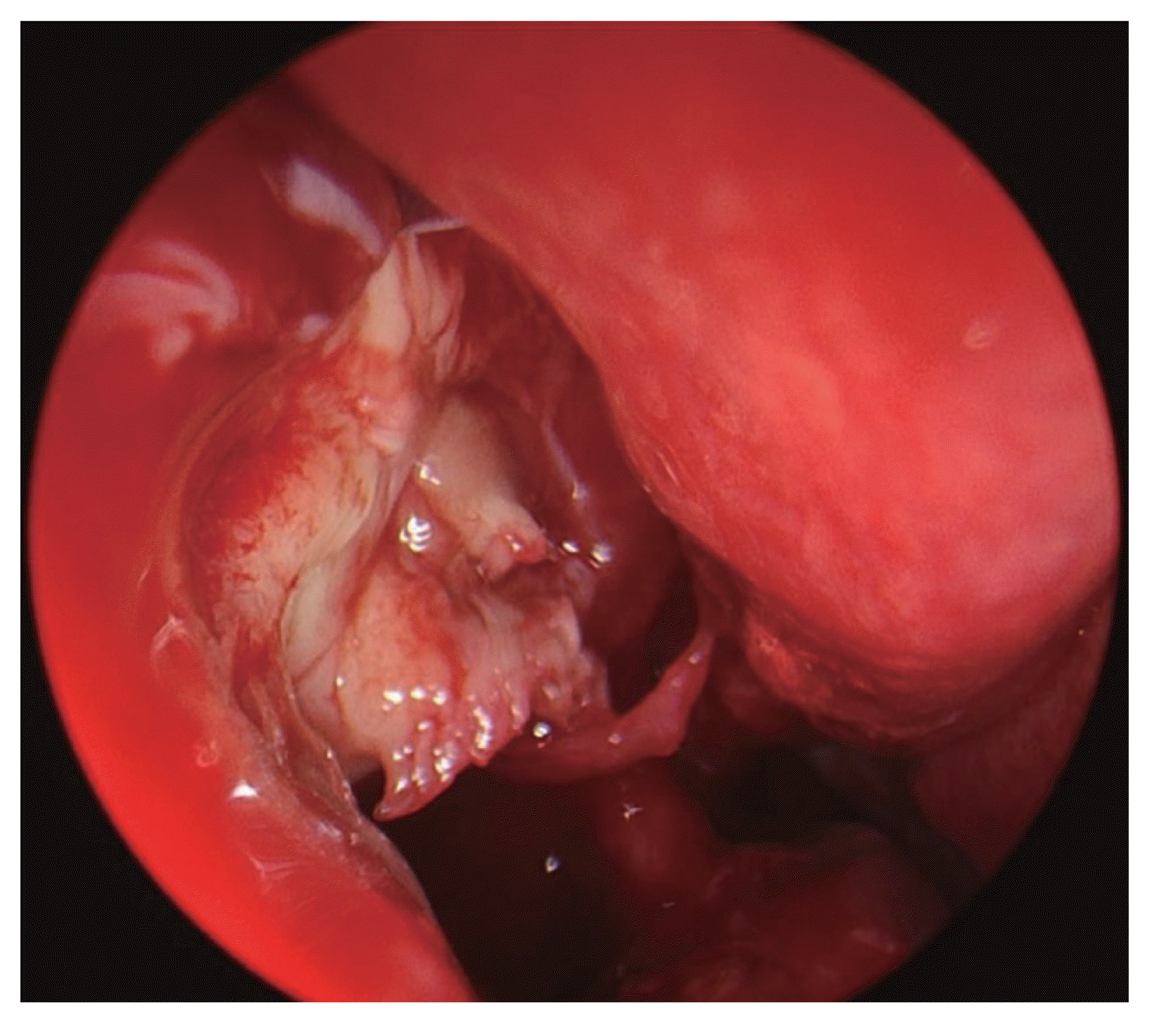
The operation was performed under general anesthesia, and when middle meatal antrostomy was performed, a mass tissue was observed in the maxillary sinus and was removed using a curved suction and forceps. The mass tissue was a soft tissue with a light yellow color (Fig. 3), since it was well separated from the surrounding tissues enough to be removed only by suction, all mass tissues visible in the surgical field were removed. When the mass tissues were removed, the suborbital wall, which showed bone erosion on CT, was exposed, and it was confirmed that the suborbital wall was well maintained without damage. In the mass tissue, yellowish-brown mucus, which is mainly seen in mucocele, was not observed. During surgery, frozen section examination was not performed. Acute complications such as epistaxis and cerebrospinal fluid were not observed after surgery, and epiphora, facial pain, and eye pain were all resolved. In histopathological examination, the cells constituting the mass were widely scattered without a certain pattern, and were partially accompanied by necrosis. When observed at high magnification, tumor cells were more than 4 times larger than the surrounding lymphocytes and had relatively abundant cytoplasm, and distinct nuclei were observed. Immunohistochemical staining showed strong positivity for the B-cell marker CD20, and Ki-67 staining showed that more than 90% of tumor cells were positive, so it was diagnosed as diffuse large B-cell lymphoma (DLBCL). It was negative for the NK cell marker CD56, so it was possible to exclude extranodal NK/T cell lymphoma (Fig. 4). In positron emission tomopraphy (PET) CT, hypermetablic mass accompanied by bone destruction was observed in the right suborbital wall (Fig. 5A), and a number of hypermetablic masses were observed in the sigmoid colon (S-colon). According to the histological examination of the hypermetabolic mass in the S-colon, it was diagnosed as a simple polyp and was thus finally diagnosed as Stage I (by Ann Arbor staging system) DLBCL confined to the right suborbital wall. One week after the surgery, the first R-CHOP (rituximab, cyclophosphamide, hydroxydaunorubicin, Oncovin, and prednisolone) chemotherapy was administered in cooperation with the hematology oncology department, and the third therapy was completed four months later, and the mucosal condition was good with no reoccurrence on nasal endoscopic examination performed at the outpatient clinic four months after the surgery. On PET-CT taken five months after the surgery, the hypermetabolism mass seen immediately after surgery was not observed and no recurrence was observed (Fig. 5B).

Pathologic findings. A: The large-sized tumor cells with vesicular nuclei showed diffuse growth pattern. Foci of cell necrosis due to rapid tumor growth were observed (hematoxylin-eosin stain, ×400). B: Tumor cells showed strong membranous positive staining for CD20 (CD20, ×400). C: Negative immunoactivity for CD56 (CD56, ×400). D: More than 90% of tumor cells were positive for Ki-67 immunohistochemical staining (Ki-67, ×400).
DISCUSSION
NHL is a type of tumor originating from the lymphoreticular system. Although NHL mainly originates in lymph nodes, 40% of cases are expressed as primary in tissues such as stomach, liver, connective tissue, meninges, and bones, which are not lymph nodes tissues. The head and neck is the second most common site following the digestive system [1]. However, in view of all malignant tumors occur in head, NHL originating from the head and neck is a very rare disease accounting for less than 1% of all malignancies. Especially, NHL originating in sinuses accounts for 0.2% of all lymphomas and accounts for only 2% in extranodal lymphoma, which emphasizes its rarity. Among the sinuses, the maxillary sinus is the most common site where the disease develops, followed by the ethmoid sinus, sphenoid sinus, and frontal sinus [2]. DLBCL is the most common subtype of NHL, and accounts for 2/3 of NHLs.
The main symptoms of NHL occurring in the sinuses are runny nose, stuffy nose, etc., and when the tumor grows and invades the orbit and palatal cavity, symptoms such as protrusion of the eyeball, double vision, and obstruction in opening mouth may occur. Since the prognosis of sinus NHL varies depending on the stage, early detection is important before the disease progresses. However, it is difficult to diagnose in early stage because the symptoms are ambiguous and nonspecific, and sometimes asymptomatic until the mass enlarges to destroy the structures of the sinus wall. Also, it is often mistaken for sinusitis or mucocele [3].
Epiphora that the patient of the case study displayed is a very rare clinical feature of sinus NHL. The main cause of epiphora is decreased tear drainage due to occlusion of the lacrimal duct and excessive tear production due to dry eye syndrome [4]. The occurrence of epiphora due to mass blocking lacrimal duct accounts for 3.8%, and among them, the case of epiphora caused by lymphoma is reported to be very rare, accounting for 0.8% of the causes for the epiphora [5].
The patient of this case developed epiphora in the right eye three months before admission and received ophthalmic treatment under the diagnosis of lacrimal duct stenosis, but did not improve, and the symptoms worsened. Dacryocystorhinostomy was planned for the patient but she developed facial pain and orbital pain, which led to further examinations to be diagnosed with maxillary sinus DLBCL.
Imaging tests such as CT and MRI are useful for diagnosing DLBCL. However, considering the characteristics of DLBCL that can show various imaging findings, histological as well as imaging tests are essential for accurate diagnosis, staging, and treatment planning. In this case as well, a mucocele was suspected by imaging findings. In general, both mucocele and lymphomas are accompanied by expansion of the bony wall during MRI and are observed as homogeneous, noncontrast-enhanced mass, showing low-intensity signals on T1-weighted images [6]. Mucoceles show high intensity on T2-weighted images, and lymphomas show mainly isointense on T2-weighted images, which makes it possible to differentiate with each other, but since lymphomas can show low to high intensity on T2-weighted images, accurate differentiation can be difficult [7]. In the patient of this case, a lesion in the maxillary sinus showing a higher intensity than that of the cerebral cortex was observed on a T2-weighted image, and it was higher intensity compared to the general lymphomas; therefore, the findings suggested more likely mucocele. However, the yellowish-brown contents observed in the mucocele were not observed in the surgery, and a pale yellow mass was observed, suggesting lymphoma. With the biopsy result, it was diagnosed as lymphoma. It is necessary to recognize the fact that lymphoma can show various imaging findings, and to comprehensively consider endoscopic findings, histology during surgery, and biopsy for an accurate diagnosis.
The pathological findings of DLBCL are characteristically large and cytoplasm-rich tumor cells compared to surrounding lymphocytes, and nucleoli are prominently observed. In addition, immunofluorescence staining was characteristically positive for the B cell marker, CD20.
The treatment for DLBCL varies depending on age, general condition, and stage, but the fundamental treatment is RCHOP, which is a combined chemotherapy, and radiation therapy. For maxillary sinus DLBCL, the main purpose of surgery is for biopsy, but it has the effect of maximally reducing tumor cells before radiation therapy to be performed later, and has the effect of alleviating the symptoms complained by the patient. Therefore, during surgery, it is necessary to reduce the size of the tumor tissue as much as possible to the extent that the surrounding tissues can be preserved and to collect sufficient specimens for biopsy [8]. For the case of this study, all tissues that were able to be observed with endoscopy during the surgery were removed, and after the surgery, the patient’s symptoms such as epiphora, facial pain, and eye pain improved immediately, and an accurate diagnosis was possible through biopsy. Complications that may occur after surgery include decreased visual acuity and facial paresthesia, and the patient complained facial paresthesia after the surgery on the side where the surgery was performed.
The probability of complete remission when R-CHOP combination chemotherapy is administered is known to be 70%, and the probability of local or cranial nerve recurrence within eight months after the chemotherapy is finished is known to be 40% [9], but the prognosis varies depending on the factors of each patient.
Although it is known that the general prognosis of DLBCL is poor with older age (over 60 years), higher lactate dehydrogenase level, higher stage, and presence of B symptoms (fever, night sweats, weight loss) [9], the prognosis of the sinus DLBCL is highly affected by the stage of the tumor, invasion into important surrounding structures, and the overall condition of the patient [10]. The patient in the case was Ann Arbor stage I, and the mass was confined to one side of maxillary sinus and nasal cavity without cranial nerve involvement. Also, considering that the patient’s general condition was good with an European Cooperative Oncology Group score of 0, the prognosis is expected to be fine.
The patient of this case started the 1st R-CHOP one week after surgery and completed the 3rd R-CHOP two months later, and five months later (as of now), the radiation treatment was all completed. On an outpatient nasal endoscopy, there was no recurrence and the mucous membrane was in good condition.
Considering that the prognosis of sinus DLBCL is stage-dependent, early detection of the disease is very important. In addition to the common symptoms of sinus DLBCL, such as runny nose, nasal congestion, post nasal drip, facial pain, and protrusion of eyes, it should be taken into account that rare clinical symptoms such as epiphora may appear as in this case. If the patient with epiphora shows no improvement with an ophthalmological treatment, but keeps worsening, doctors should suspect lacrimal duct obstruction due to mass tissue and perform image studies such as CT and MRI to diagnose in early stage of the disease. In addition, due to the characteristics of lymphoma where nonspecific imaging findings can be found, there is a possibility that it may be mistaken for a mucocele or granuloma in the interpretation of an imaging test. Therefore, sinus DLBCL should be diagnosed by comprehensively considering clinical features, endoscopic findings, and biopsy result.
Notes
Ethics Statement
Informed consent for publication was obtained from the patient.
Availability of Data and Material
The datasets generated or analyzed during the study are available from the corresponding author on reasonable request.
Conflicts of Interest
The authors have no potential conflicts of interest to disclose.
Author Contributions
Conceptualization: Young Bin Yun, Woo Yong Bae. Formal analysis: Young Bin Yun, Woo Yong Bae. Supervision: Woo Yong Bae. Visualization: Min Gyoung Pak. Writing—original draft: Young Bin Yun, Sangjun Kim. Writing— review & editing: Young Bin Yun, Woo Yong Bae.
Funding Statement
None


