A Case of Nasal Septum Gossypiboma 14 Years After Septorhinoplasty
Article information

Abstract
Gossypiboma, an infrequent surgical complication, describes a mass of cotton material inadvertently left in the body cavity after an operation. It is an extremely rare iatrogenic complication of nasal surgery, with only a few cases reported in literature to date. Here we present a case of gossypiboma in the nasal septum of a 35-year-old male patient who previously underwent septorhinoplasty fourteen years prior. He was treated by endoscopic endonasal surgery to remove the lesion. Pathologic findings showed a foreign body (gauze filament) with a giant cell reaction. This report will be helpful for treating patients with similar histories in the future.
INTRODUCTION
Residual foreign substances in the body during surgery can cause major problems, which requires caution. Nevertheless, cases of residual foreign substances in the body after surgery keep happening. Gossypiboma refers to a lesion in the form of a mass that can be formed due to cotton material left in the body during surgery. Gossypiboma occurring in the head and neck area, especially in nasal cavity, is one of the rare cases of iatrogenic complications.
This paper—along with a literature review—reports a case of gossypiboma forming in the nasal septum due to a foreign body reaction caused by remaining gauze in the nasal septum after the septorhinoplasty performed 14 years ago.
CASE REPORT
A 35-year-old male patient came to our outpatient department with persistent mucopurulent rhinorrhea, foul odor, and epistaxis in the right nasal cavity that had persisted for 1 month. The patient had no other medical history but had a history of having septorhinoplasty performed at another hospital 14 years ago. On endoscopic findings, a lesion including gauze adhered to the right septal mucosa was observed with inflammation on surrounding mucosa, and the left nasal cavity showed normal findings (Fig. 1A and B).
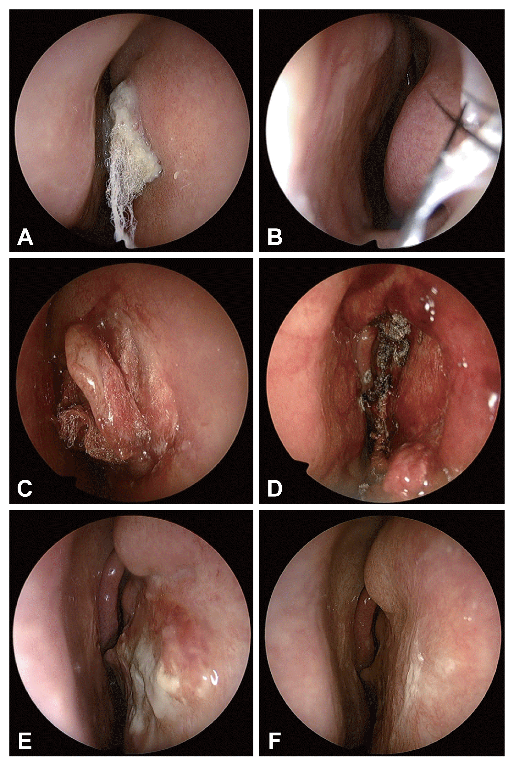
Endoscopic images of nasal cavity. A: Foreign body (gauze filament) is noted in the right nasal septum with adhesions. B: The left nasal cavity is clear. C: Foreign body (gauze filament) is imbedded in the septal cartilage with severe adhesion resulting in difficulty of removing through submucoperichondrial dissection. D: Septal cartilage was included in the total resection of the lesion. E, F: The septal mucosa is in a well-healed status without septal perforation at 1 month (E) and 3 months (F) after the surgery.
Paranasal sinuses computed tomography (CT) was performed to confirm the shape and surrounding structures of the lesion in the right nasal cavity, and a low-density infiltrating lesion was observed inside the septum in the right nasal cavity. On CT, it was difficult to accurately determine the extent of adhesion between the foreign body and cartilage, so MRI was additionally performed. On MRI, infiltrative lesions invading the right septal mucosa and septal cartilage were observed, and the left septal mucosa beyond the septal cartilage showed no adhesion. In MRI findings, the lesion was observed with low-signal intensity on both the T1-weighted and T2-weighted images, and showed no contrast enhancement on the contrast-enhanced T1-weighted image (Fig. 2).
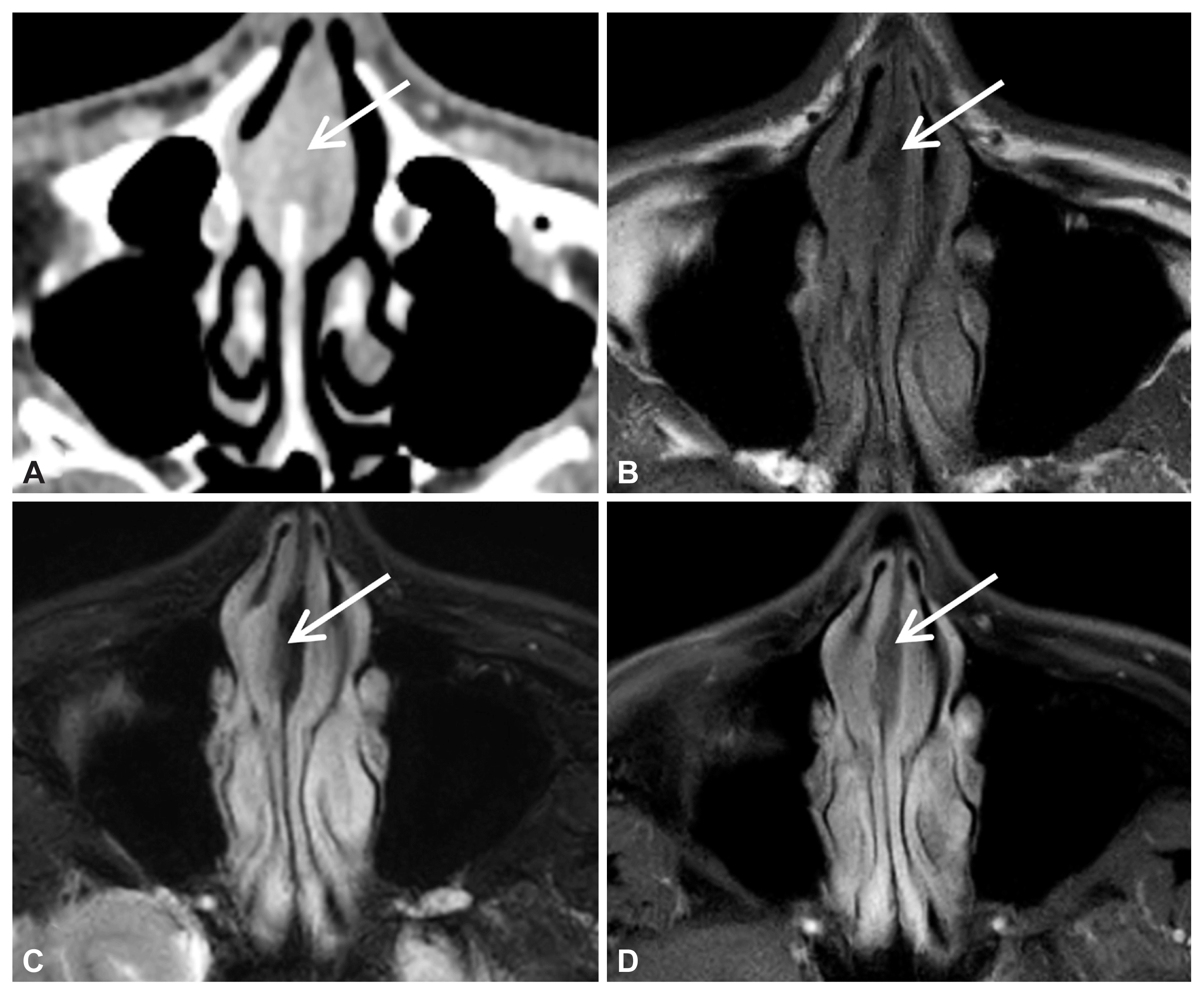
Preoperative CT and MRI findings of the lesion. CT (A) and MRI (B–D) show an infiltrative lesion in the right side of nasal septum. The lesion (arrow) shows low-signal intensity on T1-weighted image (B) and T2-weighted image (C), without enhancement on contrast-enhanced T1-weighted image (D). MRI findings show an intact mucosa and perichondrium on the contralateral side of the lesion. CT, computed tomography; MRI, magnetic resonance imaging
For treatment, a surgery to remove the entire lesion including the foreign body in the right nasal cavity using an endoscope under general anesthesia was planned. An attempt was made to remove the foreign body by preserving the septal cartilage through submucoperichondrial dissection on the right side of the nasal septum with a safety margin of 5 mm to the lesion through the right nasal cavity, but it was not easy due to a severe adhesion of the filament of the foreign body which invaded septal cartilage (Fig. 1C). Therefore, a modified Killian incision was performed on the left side of nasal septum, which is the contralateral side of the lesion, followed by submucoperichondrial dissection to completely remove the lesion, including the inflamed right septal mucosa and septal cartilage, while preserving the left septal mucosa and perichondrium (Fig. 1D). The right side of the nasal septum from which the lesion was excised was covered with gel foam after hemostasis using monopolar suction bovie. The modified Killian incision in the septal mucosa on the contralateral side of the lesion was sutured with Chromic 5-0. After inserting the silastic sheet into both nasal cavities and performing Merocel packing, the surgery was finished.
According to histopathological findings, chronic inflammatory cells, fibrosis reaction, and giant cell reaction were observed around the foreign body that looked like gauze filament, and gossypiboma was infiltrating the mucous membrane and cartilage tissue (Fig. 3). The patient had no specific findings immediately after surgery and was discharged on the 2nd day after the surgery without symptoms such as epistaxis after removing the packing. The silastic sheet was removed two weeks after surgery, and oral antibiotics of the 3rd generation cephalosporins were administered during this period. Until three months after the surgery, complications such as recurrence and septal perforation were not observed, and follow-up is being performed on an outpatient basis (Fig. 1E and F).
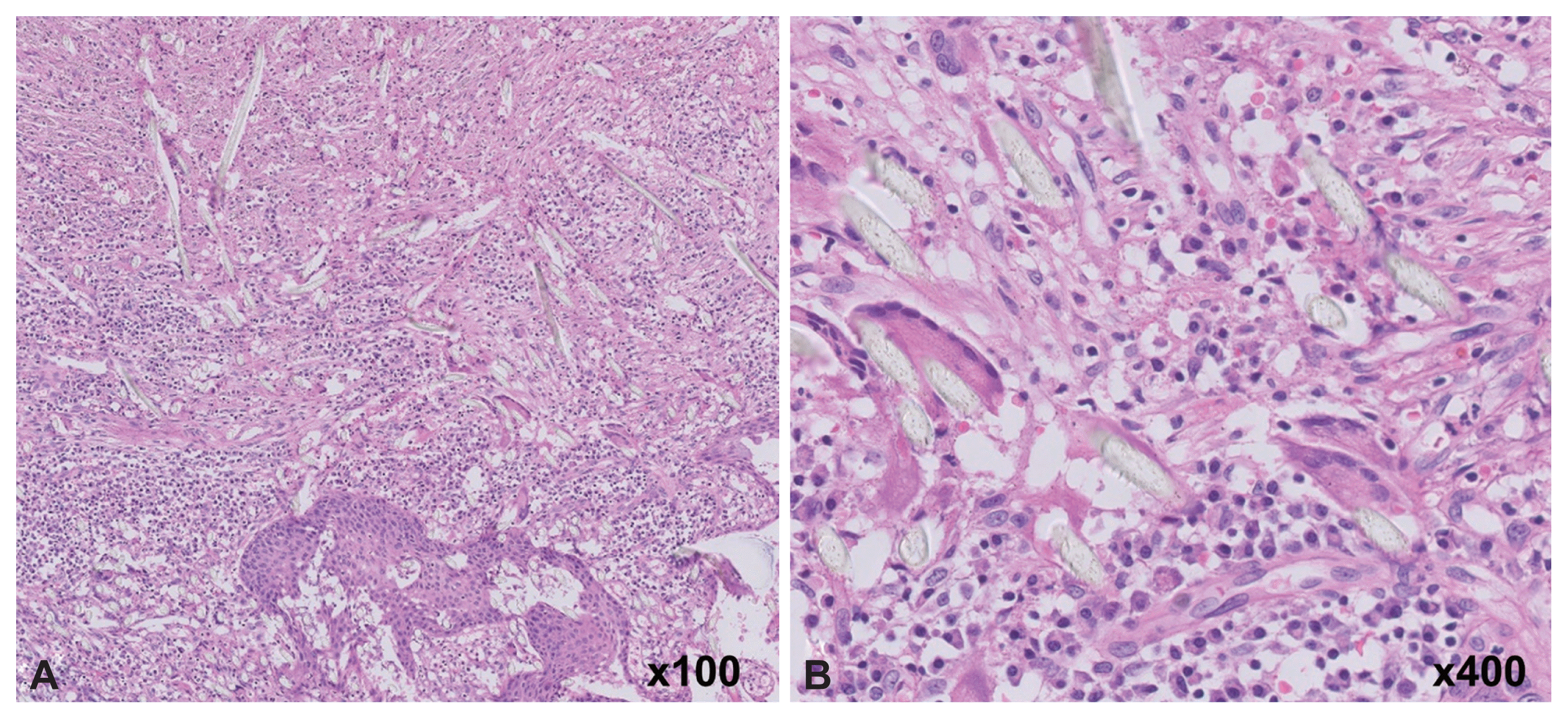
Microscopic finding. Foreign body (gauze filament) surrounded by inflammation and foreign-body giant cell reaction (hematoxylineosin, original magnification ×100 [A], ×400 [B]).
DISCUSSION
In the mentioned case, the gauze left under the nasal septum after septorhinoplasty formed severe adhesions to the septal mucosa and cartilage for 14 years, and it became a mass-shaped lesion, which ended up exposed to nasal cavity through septal mucosa causing an acute inflammatory reaction and was found due to accompanying symptoms such as mucopurulent rhinorrhea and foul odor.
Gossypiboma is a term that refers to a lesion in the form of a mass that can be formed by cotton material left during a surgery, and is one of the rare complications that can occur after a surgery. It is a complication that can occur in any surgery, and it is known that the incidence rate is higher in surgeries with a relatively wide surgical site, such as thoracotomy or laparotomy. On the other hand, case reports are rarer in the surgeries involving relatively narrow areas such as head and neckotorhinolaryngology area, especially the rhinology area [1–3]. According to one literature, the rate of gossypiboma found in the abdominal and pelvic area was 80%, while the rate found in the head and neck area was 3.96%. It took 3.35 years on average until the discovery of the disease, ranging from three days at a minimum to 35 years at a maximum [4]. According to another literature, gauze made up 69% of the foreign body left in the body after surgery and other surgical instruments accounted for 31%, and the annual incidence rate is less than 0.01% [5]. The risk factors that are highly likely to leave a foreign body in the body after surgery are cases of emergency surgeries, cases when the surgical method is changed in the middle of surgery, and cases when the patient’s body-mass index may be high. Other factors are long surgery time, multiple surgical teams involved, and change in operators in the middle of surgery [5,6].
If foreign substances remain in the body during a surgery, various complications and legal problems may occur, so it is most important to prevent it in advance. Gauze and instruments used in surgery must be thoroughly checked through standardized operational checkout procedures such as the World Health Organization Surgical Safety Checklist, and the use of gauze and instruments with radiopaque labels is recommended for prevention and diagnosis [5–7].
If foreign substances are left in the body after surgery, acute and late reactions occur. The acute reaction appears in the form of an abscess or granuloma, and the late reaction causes adhesions or encapsulation for several months to years, and various clinical symptoms associated with each reaction may occur [8]. Gossypiboma should be differentiated from an abscess at the surgical site, other benign tumors and malignant tumors, and for this, a detailed history taking, physical examination, and appropriate imaging and biopsy are required [9].
Gauze with radiopaque labels or radiopaque surgical instruments can be easily detected with simple radiographs, but otherwise, there is a limit to the diagnosis of gossypiboma. CT is useful in diagnosing gossypiboma and its associated complications. A low-density lesion formed from gauze is seen on CT, and a sponge pattern of air bubble shading is shown inside, and a film with enhanced contrast can often be seen around it [10]. Occasionally, calcification can be seen on CT, and remodeling of surrounding tissues can also be observed due to lesions [8,10]. In MRI, gossypiboma show various signal intensities depending on the water and protein components inside the lesion, and most of them show low-signal intensity on T1 image and variable signal intensities on T2 image [8,10]. Ultrasound can be used for other imaging tests, but there is a limit to the test in other otorhinolaryngology areas except for the head and neck area. The case was observed as a low-density shading lesion on CT, and both T1-weighted and T2-weighted images showed low-signal intensity on MRI.
Giant cell reaction and fibrotic reaction are characteristic findings that can be caused by the chronic inflammatory reaction of a foreign body [11]. The same findings were observed in the histopathologic findings of this case. In addition, the gossypiboma formed by a chronic inflammatory reaction was infiltrating the surrounding mucosa and cartilage tissue. These findings indicate that a chronic inflammatory reaction occurred around the gauze filament existing in the sterile space between the septal cartilage and perichondrium, slowly infiltrating the surrounding tissues, and then exposed to the outside, causing an acute inflammatory reaction.
If gossypiboma is diagnosed, the previous surgical site should be reopened immediately to remove the remaining gauze and surrounding inflammatory tissue and granulation tissue formed. In addition, it should be accompanied by appropriate antibiotic treatment through bacterial culture test, and depending on the situation, it may be necessary to insert a drainage tube. In this case, as well, after the diagnosis of gossypiboma, surgery was performed to remove the surrounding inflamed tissue, including the gauze, as soon as possible. Since there was no adhesion in the mucosa on the contralateral side of the lesion on MRI, endoscopic endonasal surgery was attempted. If an adhesion on the contralateral side was observed, external nasal approach to remove the lesion would have been considered.
This case is significant in that it was reported in the otorhinolaryngology department, where the incidence of gossypiboma is very low, and among them, the rhinology department. A case in which gossypiboma progressed as the remaining gauze in the nasal septum gradually adhered to the surrounding mucosa and cartilage for a long time after septorhinoplasty 14 years ago is difficult to find in existing literature reports. The reason the patient was asymptomatic for 14 years was that the residual gauze was placed in an aseptic state between the relatively hard septal cartilage and the perichondrium, so adhesions and granulation tissue formation reaction took place gradually around the periphery over a long period of time. It seems that the patient’s symptoms appeared by an acute inflammatory reaction caused by the gauze is exposed to the nasal cavity due to weakened septal perichondrium mucosa.
In this case, by performing preoperative CT as well as MRI, it was confirmed in advance that the lesion did not adhere to the contralateral septal mucosa. Therefore, among two approaches—endoscopic endonasal surgery and external nasal approach—less invasive endoscopic approach was able to be adopted safely. In addition, submucoperichondrial dissection was performed to preserve the septal mucosa and perichondrium on the contralateral side of the lesion thereby reducing the possibility of postoperative septal perforation, not just impractically excising the entire lesion with severe adhesion to the surrounding tissue only through the lesion side approach. It is expected that the examination and treatment experience performed in this case will be helpful when facing patients with similar medical histories in the future.
Notes
Ethics Statement
This study was reviewed and approved by the Institutional Review Board of Yongin Severance Hospital (IRB No. 4-2020-1154), and informed consent was waived.
Availability of Data and Material
The datasets generated or analyzed during the study are available from the corresponding author on reasonable request.
Conflicts of Interest
The authors have no potential conflicts of interest to disclose.
Author Contributions
Conceptualization: Jong-Gyun Ha. Data curation: Seungjoon Yang. Funding acquisition: Jong-Gyun Ha. Methodology: Sohi Bae, Jong-Gyun Ha. Visualization: Seungjoon Yang, Sohi Bae, Jayeong Seo. Writing—original draft: Seungjoon Yang. Writing—review & editing: Sohi Bae, Jayeong Seo, Hyung-Ju Cho, Chang-Hoon Kim, Jong-Gyun Ha.
Funding Statement
This study was supported by a faculty research grant of Yonsei University College of Medicine for 6-2020-0118.