Computed Tomography and Anatomical Findings Encountered During Revision Endoscopic Sinus Surgery
Article information
Abstract
Background and Objectives
Functional endoscopic sinus surgery (FESS) is a well-established strategy for the treatment of rhinosinusitis. However, some patients do not respond to primary surgery and may require revision surgery. Anatomic alterations due to prior sinus surgery, scarring and adhesions as well as associated chronic mucosal inflammation can make revision procedures challenging. In order to shed more light on the difficulties faced by surgeons performing revision FESS, a study was performed to identify areas of recurrent disease on computed tomography in patients undergoing revision surgery, as well as to evaluate intraoperative findings during revision FESS.
Materials and Method
A hospital-based, interventional, non-randomized study was undertaken in 40 patients who underwent revision FESS. Multiple clinical parameters were recorded including number and type of previous surgeries, latest CT scans of the nose and paranasal sinuses, as well as intraoperative findings.
Results
Our findings demonstrated the diffuse nature of mucosal disease on CT in our patient population. Fibrosis and adhesion formation were the most common intraoperative findings on revision sinus surgery along with residual air cells, polypoid mucosal regrowth, and middle meatal antrostomy stenosis.
Conclusion
A careful evaluation of the patient is needed while contemplating revision surgery. A recent high-resolution CT scan is of paramount importance. The most common areas of disease recurrence are the ostiomeatal complex and residual ethmoids, and these areas should be given careful attention.
INTRODUCTION
Chronic rhinosinusitis today continues to be a significant cause of morbidity all over the world. The unpredictability of the disease often coupled with fluctuations in relief and worsening of symptoms of patients is a point of concern for rhinologists, who still face this dilemma in their daily practise.
Functional endoscopic sinus surgery (FESS) has over time become the established surgical treatment for chronic rhinosinusitis cases which do not respond to medical management [1]. Multiple studies conducted over decades have shown success rates for FESS as high as 98% if done properly [2]. However, a group of patients still exist who do not have a continued benefit from FESS with persistent or recurrent symptoms, and these patient often require revision surgery [3]. But there are several factors that make a revision surgery for chronic rhinosinusitis challenging for the operating surgeon such as several pathological changes, recurrent polyps in the nose, hyperostotic ethmoid cells, as well as altered anatomy due to previous surgery, mucosal scarring and associated chronic mucosal inflammation. Therefore, revision surgeries for chronic rhinosinusitis pose a major complexity even with most experienced endoscopic surgeons with high chances of failure or negative outcomes. One of the key steps in revision surgery is identifying the altered anatomy and resulting pathology that leads to recurrence of disease. Once identified, a thorough and complete removal of all diseased tissue remains the cornerstone for achieving successful results in revision surgeries [4].
In order to shed more light on the continued difficulties faced by surgeons performing revision surgeries especially when they lack access to intraoperative neuroimaging, a study was performed to identify the areas of recurrent or persistent sinonasal disease in patients undergoing revision endoscopic sinus surgery using computed tomography findings, as well as to evaluate the intra-operative findings including anatomical changes following previous surgery that may be encountered while performing revision endoscopic sinus surgery.
MATERIALS AND METHODS
A hospital-based, interventional, non-randomized, non-comparative and prospective study was undertaken in which the outcomes of 40 patients who had undergone Revision Endoscopic sinus surgery were reviewed. Study duration was from June 2017 to June 2019. An informed consent was obtained for each patient, as well as institutional ethical committee clearance was secured at the start of the study.
Selection criterion: presence of recurrent/persistent symptoms despite maximal medical therapy.
Exclusion criteria: Pregnant females and any patients below 12 years (paediatric age group).
Multiple clinical parameters were recorded for each patient including age, gender, symptoms, details of previous surgery, previous Computed Tomography (CT) of nose and paranasal sinuses, and diagnostic nasal endoscopy was done.
Prior to their Revision Endoscopic Sinus Surgery (RESS) procedures, all patients were treated with recommended courses of antibiotics for a period of four to six weeks, and most required course of oral and nasal steroid therapy. Decongestants were used in initial phase of therapy to alleviate symptoms.
High resolution Computed tomography (CT) with 1 mm cuts was performed one month before all revision surgeries while patients were undergoing initial medical management. The mucosa of all sinuses including the ostiomeatal complex status was studied as per the Lund-Mackay system [5] and were scored accordingly i.e a sinus with no opacification (score 0), a sinus showing partial opacification (score 1) and a sinus showing full opacification (score 2).
All 40 patients with 79 operative sides underwent a complete spheno-ethmoidectomy with/without endoscopic assisted polypectomy. Special attention was given to use mucosal sparing instruments and powered instrumentation like microdebrider where required. Postoperatively, all patients were given a course of antibiotics, nasal decongestants, nasal irrigations, intranasal steroids and oral steroids (if associated with nasal polyps) for a minimum period of 4-6 weeks.
The study included the patients from all age groups and of both the sexes.
The data was tabulated on presence or absence of recurrent chronic rhinosinusitis with/without sinonasal polyposis, the significant pathological factors that contributed to the recurrence of pathology, as well as the change in anatomy seen while performing revision surgery.
RESULTS
In our study, 80 patients out of total 200 (40%) who had previously undergone endoscopic sinus surgery, presented with persistent recurrent symptoms, out of which 40 patients were successfully managed with medical therapy alone (20%) whereas the remaining 40 patients (20%) required a revision sinus surgery. Therefore, a total of 40 patients with 79 operated sides were assessed in this study.
Of those 40 patients aged 19 to 64 years, 23(67.5%) were males and 17(42.5%) were females. Revision sinus in our study was done bilaterally for 97.5% of cases and unilaterally for 2.5% cases. The interval between previous surgery and onset of recurrent symptoms ranged from less than 1 year to more than 5 years in our study with duration of recurrent symptoms after previous surgery ranging from 7 months to 8 years. Multiple symptoms were reported by most patients, the most commonly occurring symptom was nasal obstruction (30 patients, 75%) followed by olfactory disturbances (22 patients, 55%), nasal discharge (18 patients, 45%), and headache and recurrent infections (17 patients, 42.5%). Out of 40 patients in our study, 36 (90%) has a history of a single previous endoscopic sinus surgery, 3 (7.5%) had a history of previous two surgeries, and 1(2.5%) had a history of three prior surgeries.
Mucosal status of sinuses on CT scans as graded by Lund Mackay system are shown in Table 1.

Mucosal status of sinuses on CT scan
The incidence of anatomic abnormalities and the intraoperative findings have been shown in Table 2 and 3 respectively.
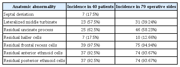
Anatomical abnormalities encountered during nasal endoscopy

Intraoperative findings during revision sinus surgery
DISCUSSION
The advances and increased accuracy of CT scans along with higher quality endoscopic visualization with newer camera and imaging systems have vastly aided rhinologist in identifying the anatomy of sinus with its variations as well as explore the pathogenesis contributing to the elusive nature of chronic rhinosinusitis. The role of anterior ethmoid sinus and ostiomeatal complex in primary as well as recurrent disease has time reiterated time and again by surgeons. In addition to the above, the concept of mucosal disease and its subjective nature with different patients is continuously studied to improve the surgical outcomes for chronic rhinosinusitis.
The distribution of disease in our study as seen by the mucosal status on CT scans was assessed by studying involvement five specific areas in 79 operated sides which showed anterior ethmoid disease in 93.67 %, maxillary sinus disease in 96.2%, and frontal sinus disease in 78.48%. Posterior ethmoid and sphenoid sinus involvement were noted in 93.67% and 72.15% of the 79 operated sides. This clearly demonstrates the diffuse character of mucosal pathology in our study population. Over the past two decades, many studies have shown the role of osteomeatal complex in the pathogenesis of recurrent disease [6,7] which forces us to explore this complex again during revision surgery, and points to the fact that paying attention to these areas in the primary surgery itself may perhaps help avoid revision surgeries to begin with. However, the widespread multisinus disease noted in our study suggests that a factor in addition to obstruction of the ostiomeatal complex is involved in the pathogenesis of chronic sinusitis in these patients. As noted by Lawson and Kennedy [8,9], there are several additional factors that contribute to the mucosal response and lead to persistent disease progression.
During our literature review, we found that not many studies spoke in detail about the anatomic changes and findings encountered while performing revision FESS. In one prominent study the authors clearly stated that the most common anatomy finding they saw was lateralisation of the middle turbinate, followed by residual anterior ethmoidal cells followed by scarred frontal recesses [2]. In comparison, in our current study we noticed a high incidence of residual cells (Table 2). We found that 93.67% of the studied sides (92.5% patients) had both residual anterior as well as posterior ethmoidal cells, and 94.94% of the sides (97.5% patients) had residual frontal recess cells. In contrast, lateralization of middle turbinate was noted only in 39.24% sides. Our assessment is that this may be due to more conservative or limited FESS techniques which may be followed by lesser experienced or younger surgeons. The counter argument in FESS as a conservative procedure has always been that removal of all sinus cells may not be necessary in most cases, and the decision to limit surgery or do a more widespread removal should be made as per each individual patient. However, in our study, most of the patients had pansinusitis, as per the CT scores, where only 3.8% of maxillary sinuses, 6.33% of anterior and posterior ethmoid sinuses whereas only 21.52% of frontal sinuses showed no opacification. We therefore found it reasonable to assume that most of the patients perhaps required more thorough and complete surgical than what was done during the initial procedures.
During the revision surgery, we carefully recorded the findings (Table 3) that we considered were contributing to the recurrence of the disease. Intra-operative findings in our study were noted for 79 sides while performing RESS on each side.
Our study showed that incidence of residual uncinated process was 58.23% (62.5% of the patients). This finding was consistent with studies that advised identifying a remnant of uncinate process as a crucial step while performing revision surgery [4].
Residual anterior and posterior ethmoid cells were noted in 70 (88.61%) and 73 (92.41%) operative sides respectively. The sphenoid sinus ostium was obstructed in 52 (65.82%) sides either due to polyps/polypoidal mucosa or scarring due to previous surgery. Scarring after previous surgery in the middle meatus was seen in 11 (13.92%), and adhesions of middle turbinate was seen in 6 (7.59%) of these cases. Frontal recess scarring was noted in 2(2.53%) of operative sides. In addition, lateralized middle turbinate was seen in 31 (39.24%) operated sides without adhesions. Out of 79 operated sides, 29 (36.71%) showed signs of middle turbinate dissection with broken ground lamella observed in 34 (43.04%) of the operated sides. Frontal sinus outflow obstructed by agger nasi cells was seen in 5 (6.33%) sides. Presence of polyps or polypoid mucosa was observed in 55 of 79 (69.62%) sides.
Our findings were consistent with established studies that found fibrosis and adhesion formation was the most consistent and recurring sign noted in revision surgeries [10], while other significant contributing factors included residual air cells, polypoidal mucosa, and stenosis of middle meatus [11,12]. These findings reiterated the findings of the CT scans, and we as surgeons were forced to consider that perhaps a lot of these revision cases could be avoided if careful attention was perhaps given during the primary surgery and consistent follow-up after initial surgery.
Endoscopic evaluation 6 months postoperatively found the majority of patients had clear sinus cavities and the mucosa of sinuses recovered. Nevertheless, 10 patients (25%) had mucosal edema, 5 patients (12.5%) had nasal discharge, 1 patient (2.5%) developed adhesions between inferior turbinate and septum. No adhesions were seen between the lateral nasal wall and middle turbinate. Also noted were polyps in 3 patients (7.5%) and crusting in 2 patients (5%). Symptoms of nasal obstruction and persistent nasal discharge seen in our patients responded well by the 6 month postoperative period. Check endoscopy done in the postoperative period confirmed a lower incidence of recurrent disease as compared to previous surgeries, but mucosal edema was not rare.
CONCLUSION
Although FESS helps more than 80% of patients in improving their symptoms, a set of patients do require revision surgery in due course. Many factors contribute to recurrent or persistent disease such as patient factors (difficult anatomy, mucosal factors), disease factors (nasal polyposis, allergic sinusitis), surgeons’ inability to deal with the disease, or poor postoperative healing. Careful attention and timely anticipation to these factors might help to prevent failure of surgery.
An even more careful evaluation of the patient is needed while contemplating revision surgery. A thorough nasal endoscopy of sinonasal cavity as well as a fresh high resolution CT scan is of paramount importance. The surgery, when indicated, is tailored according to the disease and anatomical findings. The most common areas of disease recurrence are ostiomeatal complex and residual ethmoids, and these areas require special attention. All the ethmoid partitions should be removed and a complete spheno-ethmoidectomy achieved might help ensure maximal surgical outcome.
Finally, if the common sites of recurrence are paid a thorough attention during the primary surgery itself, it may forego the need of a revision surgery at all.
Acknowledgements
We would like to acknowledge the doctors and nursing staff in our department who participated in the treatment of these patients.