Isolated Sphenoid Fungal Sinusitis With Mucocele Mistaken for Chordoma: A Study of Two Unique Cases
Article information

Abstract
Isolated sphenoid fungal sinusitis (ISFS) is a rare condition characterized by fungal infection of the sphenoid sinus. It often presents with non-specific symptoms, which can lead to misdiagnosis. This study presents two unique cases of ISFS with mucocele that were initially misdiagnosed as chordoma based on preoperative radiographic findings. Two cases of ISFS were thoroughly investigated, including clinical examinations, radiological assessments, and surgical explorations. The patients’ symptoms, radiographic findings, surgical procedures, and postoperative outcomes were documented. In both cases, radiographic assessments raised suspicion of chordoma due to bony destruction and soft tissue lesions involving the sphenoid sinus and clivus. However, endoscopic sinus surgery revealed fungal balls and mucoceles, confirming the diagnosis of ISFS. Postoperative pathology confirmed the presence of aspergilloma. The patients recovered well with appropriate treatment. ISFS is challenging to diagnose due to its deep anatomical location and non-specific symptoms. Visual disturbances, particularly affecting the abducens nerve, are common. Sphenoid sinus mucocele, though rare, can be present. Surgical exploration plays a crucial role in establishing an accurate diagnosis and initiating appropriate treatment. ISFS can mimic other skull base lesions, such as chordoma, on preoperative radiography. These cases underscore the value of surgical exploration in reaching an accurate diagnosis and highlight the need for the cautious interpretation of radiological findings in sphenoid sinus lesions to ensure optimal patient care.
INTRODUCTION
Isolated sphenoid fungal sinusitis (ISFS) is a rare entity characterized by fungal infection of the sphenoid sinus, with no involvement of the other paranasal sinuses. It often presents with non-specific symptoms, making it challenging to diagnose accurately [1]. This paper describes two unique cases of ISFS with mucocele that were initially misdiagnosed as chordoma based on preoperative radiographic findings. These cases highlight the importance of distinguishing between these two conditions and emphasize the need for thorough evaluation and surgical exploration to arrive at an accurate final diagnosis.
CASE REPORTS
Case 1
A 53-year-old man presented to the otorhinolaryngology outpatient clinic with sudden-onset diplopia that occurred exclusively when looking to the left. On examination, the left eye exhibited restricted lateral gaze, while the right eye had normal mobility (Fig. 1). Radiographic investigations, including paranasal sinus (PNS) computed tomography (CT) and brain magnetic resonance imaging (MRI), raised concern for a lesion involving the left sphenoid sinus, with potential extension into the upper clivus. The radiologist described the findings as perplexing, which suggested the possibility of a skull base chordoma or chronic sinusitis with mucocele. However, the presence of heterogeneous enhancements accompanied by bone destruction in the upper clivus raised concerns about the possibility of a skull base chordoma. Therefore, pathological confirmation under operation was recommended for the central lesion in the upper clivus (Figs. 2 and 3).
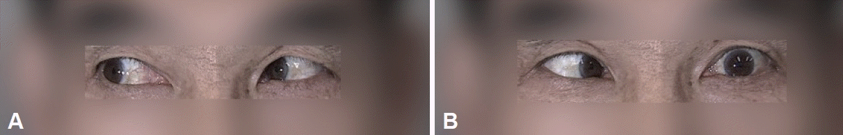
Preoperative extraocular movement assessment (Case 1). A: When the patient looks to the right, both eyes normally gaze to the right. B: When the patient looks to the left, the right eye normally gazes to the left, but the left eye cannot.

Paranasal sinus (PNS) computed tomography (CT) findings (Case 1). A: PNS CT axial view showing soft tissue density within the left sphenoid sinus and focal bone dehiscence of the lateral wall and posterior wall. B: PNS CT sagittal view showing that the lesion in the sphenoid sinus is connected to the upper clivus through the focal bone dehiscence.

Brain magnetic resonance imaging (MRI) findings (Case 1). A: T1-weighted brain MRI showing high signal intensity within the sphenoid sinus. B: T2-weighted brain MRI showing low signal intensity within the sphenoid sinus. C: T1-weighted brain MRI showing no enhancement upon contrast.
Endoscopic sinus surgery (ESS) was carried out to explore the inside and surroundings of the left sphenoid sinus. Upon surgical exploration, a fungal ball was discovered within the left sphenoid sinus, along with a mucocele situated behind it (Fig. 4). It is presumed that the fungal ball, which blocked the sphenoid sinus orifice and formed a mucocele inside the sinus, resulted in skull base erosion, which was mistaken for chordoma on brain MRI. Contrary to the preoperative imaging, neither chordoma nor ecchordosis physaliphora was found during the procedure. The patient’s diplopia completely resolved on the fourth day post-surgery (Fig. 5). Based on the surgical findings, after removing the fungal ball within the left sphenoid sinus, there was pus drainage from a small defect in the skull base, raising suspicion of fungal infection extending into the cavernous sinus. Postoperatively, the surgical findings were immediately shared with an infectious disease specialist for consultation regarding further treatment. In accordance with their recommendation, the patient received intravenous ceftriaxone and amphotericin B until the fifth day post-surgery. Postoperative pathology confirmed the presence of aspergilloma. The patient completely recovered and was discharged.

The fungal ball is being removed through the sphenoid sinus orifice (Case 1).
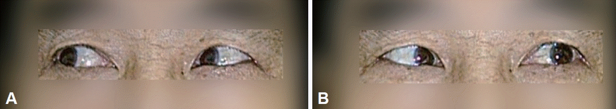
Postoperative extraocular movement assessment (Case 1). A: When the patient looks to the right, both eyes normally gaze to the right. B: When the patient looks to the left, both eyes normally gaze to the left (Improved eye movement compared to the preoperative status).
Case 2
A 62-year-old man experienced sudden-onset double vision while driving; it worsened upon rightward gaze but disappeared when looking to the left. Upon presenting to our hospital, the patient was initially admitted to the neurology department, where the double vision spontaneously resolved after 7 days. Radiographic assessments were carried out. A PNS CT scan showed a 3.0-cm lobulating cystic tumor with a bony defect extending into the left sphenoid sinus, filled with fluid (Fig. 6). Additionally, there was a wide defect in the posterior cortex of the clivus with peripheral sclerotic bone changes. Based on PNS CT, the initial differential diagnosis identified mucocele as the most likely possibility, while empyema or fungal sinusitis were considered less likely. Brain MRI displayed a maximum 3.2-cm expansile lesion that was centered on the clivus, predominantly involving the sphenoid region (Fig. 7). The initial differential diagnosis included chordoma, aneurysmal bony cyst, or sphenoid mucocele. After thorough imaging examinations, lesions requiring surgical treatment were confirmed. For surgical evacuation, he was transferred to the otorhinolaryngology department.

Paranasal sinus (PNS) computed tomography (CT) findings (Case 2). A: PNS CT axial view, showing an approximately 3.0-cm lobulating bone defect continuing to the left sphenoid sinus and the wide defect in the posterior cortex of the clivus. B: PNS CT sagittal view showing a wide bone defect between left sphenoid sinus and clivus.

Brain magnetic resonance imaging (MRI) findings (Case 2). A: T2-weighted brain MRI, showing high signal intensity within the sphenoid sinus. The sphenoid sinus and clivus lesion are distinguished by a layer of mucosa. B: T1-weighted brain MRI showing low signal intensity within the sphenoid sinus. C: T1-weighted brain MRI showing no enhancement upon contrast. Only a layer of mucosa showed rim enhancement.
ESS was performed, leading to the discovery and removal of a fungal ball within the left sphenoid sinus, along with the mucocele behind it. It is suspected that a fungal ball obstructed the left sphenoid sinus orifice, leading to the formation of a mucocele inside the sinus, which in turn caused skull base erosion. The above-mentioned bone erosion is suspected to have created the possibility of a misdiagnosis as chordoma on preoperative brain MRI. Contrary to the preoperative brain MRI, neither chordoma nor aneurysmal bony cyst was identified during surgery. Postoperative pathology confirmed the presence of aspergilloma. The patient recovered well and was discharged as scheduled.
DISCUSSION
Historically, delayed diagnosis and treatment of sphenoid sinus opacification have led to high mortality rates [2,3]. Diagnosing and treating ISFS accurately is a challenging task, but considering the impact and distress it can cause to patients, the role of a physician is crucial. Due to its deep anatomical location, a thorough diagnosis of ISFS can only be achieved by employing both clinical and radiological assessment methods. Therefore, it is imperative for physicians to remain watchful, as even a slight lapse in attention could lead to a missed diagnosis and hinder the achievement of treatment goals for this condition.
These cases described herein highlight the diagnostic challenges associated with ISFS, which can mimic other pathologies such as chordoma on preoperative imaging [4-6]. Mucocele and fungal ball commonly exhibit low signal intensity on both T1- and T2-weighted imaging. In the mucosal membrane of both disease entities, an inflammatory reaction can occur, which may manifest as thin peripheral enhancement [7,8]. The majority of sinonasal tumors exhibit a T1 signal ranging from hypointense to isointense when compared with the muscle tissue. Furthermore, these sinonasal neoplasms often demonstrate uniform or varied enhancement. The specific manner in which these tumors enhance on contrast imaging is crucial for differentiating them from neoplasms, as inflammatory conditions typically show a distinct pattern of thin and intense enhancement around the periphery [9,10]. In both cases, the preoperative imaging study raised suspicion of chordoma due to erosive bony destruction and soft tissue lesions involving the sphenoid sinus and clivus. Upon retrospective analysis post-treatment, it appears that both cases warranted a surgical approach, considering the possibility of either a mucocele or a fungal ball. This assessment was based on MRI findings where the main body of the tumor did not demonstrate distinct contrast enhancement, yet there was notable enhancement in the peripheral regions. Fungal balls in the sphenoid sinus pose a distinctive challenge because they have the potential to gradually lead to localized erosion [11]. However, ESS finally revealed fungal balls and mucoceles, ultimately confirming the diagnosis of ISFS. The distinction between these disease entities is important due to differences in treatment and prognosis.
The most frequently observed symptom of ISFS is a persistent headache that does not respond well to medical treatment. ISFS can manifest not only as headaches, but also as visual disturbances like decreased visual clarity, hazy vision, and cranial nerve dysfunction [6,12-14]. This underscores the challenges of diagnosing sphenoid sinusitis, which can be attributed to its gradual onset and the non-specific nature of headaches related to its location. Among the cranial nerves, the optic nerve and the abducens nerve are frequently affected, likely due to their proximity to an anatomically deep position within the cavernous sinus, making them susceptible to various medical issues [6].
Sphenoid sinus mucocele is a rare condition, accounting for only 1%–3% of all mucoceles within the entire nasal cavity [3,14,15]. In most cases, it is predominantly found in the ethmoid and frontal sinuses. The etiology is still unclear; however, it is suspected to be related to trauma or infective factors [15]. In the two cases presented in this report, it is postulated that a fungal ball obstructed the orifice of the sphenoid sinus, leading to the formation of a mucocele within the sphenoid sinus.
These cases emphasize the need for a comprehensive diagnostic approach, including surgical exploration, when confronted with atypical presentations, as preoperative radiographic assessments alone may not provide a clear and definite diagnosis. When ESS is performed with an uncertain diagnosis, it is a sufficiently safe and effective therapeutic approach for diverse sphenoid lesions [3,12,14]. Furthermore, ISFS with mucocele should be considered in the differential diagnosis of sphenoid sinus lesions, particularly when imaging suggests chordoma or another aggressive lesion. Clinicians, including otorhinolaryngologists, should remain alert and open to the possibility of rare and unexpected pathologies. These cases emphasize the need for cautious interpretation of radiographic findings and highlight the clinical utility of ESS in establishing the correct diagnosis.
In conclusion, ISFS can present with radiographic features that mimic other skull base lesions, such as chordoma. These cases demonstrate the value of surgical exploration in establishing an accurate diagnosis and initiating appropriate treatment. Clinicians should remain vigilant when interpreting radiological findings in cases of sphenoid sinus lesions to prevent misdiagnosis and ensure optimal patient care.
Notes
Ethics Statement
This case report was approved as exempt and the waiver of patient consent was obtained from the Institutional Review Board of Korea University Ansan Hospital (exemption number 2023AS0283).
Availability of Data and Material
All data generated or analyzed during the study are included in this published article.
Conflicts of Interest
Min Young Seo who is on the editorial board of the Journal of Rhinology was not involved in the editorial evaluation or decision to publish this article. The remaining author has declared no conflicts of interest.
Author Contributions
Conceptualization: Min Young Seo. Funding acquisition: Min Young Seo. Investigation: Min Young Seo, Kukjin Nam. Project administration: Min Young Seo. Supervision: Min Young Seo. Writing—original draft: Kukjin Nam. Writing—review & editing: Min Young Seo.
Funding Statement
This study was funded by Korea University Ansal Hospital Grant(O2310621).
Acknowledgements
None