Perimenstrual Epistaxis Caused by Nasal Septal Endometriosis: Very Rare Case Report
Article information

Abstract
We present a case of localized nasal endometriosis to contribute to the literature. A primiparous, primigravida, 37-year-old female was diagnosed with nasal septal endometriosis in the presence of history of periodic epistaxis, nasal pain, and nasal fullness. After a nasal biopsy for exclusion of malignancy, a comprehensive general screening was performed on the recommendation of a gynecologist, definitive endometriosis was diagnosed, and oral treatment was initiated. Significant relief was reported in her nasal symptoms during the 1-year follow-up period. We report this case to demonstrate the importance of clinical awareness of periodic cyclic perimenstrual epistaxis, nasal lesions, and nasal pain as a cause of nasal septal endometriosis.
INTRODUCTION
Endometriosis is a common chronic inflammatory condition gynecological disease with the presence of endometrial stroma and glands outside the uterine cavity. Extrapelvic endometriosis has been described in almost every organ. The gastrointestinal and urinary tracts are the most common sites of extrapelvic endometriosis. Nasal septal mucosal localized endometriosis is very rare; there are only two cases in the literature. We would like to present you highly exceptional nasal localized endometriosis case report to contribute to the literature. Clinicians should have to know that periodic cyclic perimenstrual epistaxis, nasal lesions, and nasal pain, which may rarely be the cause of the nasal septal endometriosis.
CASE REPORT
A primiparous, primigravida 37-year-old female comes to the ENT outpatient clinic cause of perimenstrual epistaxis, wound, nasal fullness, and pain for about one year. While an endoscopic nasal examination was performed through the left nostril, a fragile, crusted, erosive lesion was observed in the anterior of the nasal septum (Fig. 1A and B). Examination of the right nasal cavity and nasopharynx was normal. Paranasal CT and facial-nasopharyngeal MRI were performed to rule out malignancy and other pathological conditions (Fig. 1C and D). The report resulted in a 1×1 cm ulcerative-granulative lesion that does not invade the septal cartilage and only erodes the mucosa. After that was decided to take a biopsy from the lesion (Fig. 2A). Erosive tissue in the left nasal septum mucosa, which did not invade the cartilage, was excised in the subperitoneal plane after the cauterization of minor submucosal bleeding used absorbable intranasal sponges with antibiotic creams. The histologic study revealed the appearance of more than one islet-shaped stroma and glandular structures from nasal septal lesion biopsy.
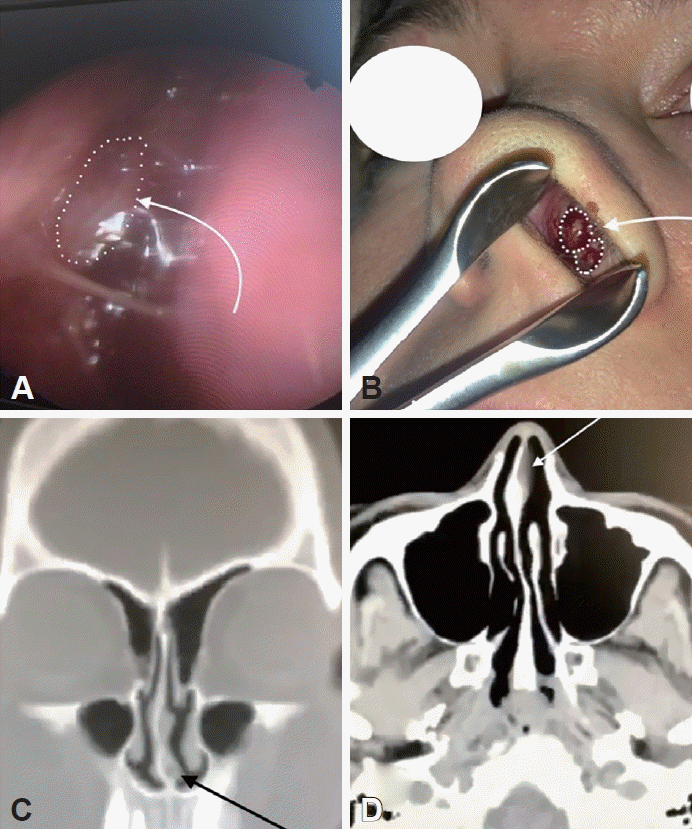
A: Left nostril endoscopic view. B: Left nostril anterior rinoscopy. C: Facial MR views: Coronal profile. D: Facial MR views: Axial profile. Nasal mucosal lesions shown with white dots and arrows.
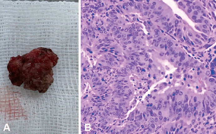
A: Macroscopic view of biopsy material from nasal mucosa (1×1 cm). B: Histopatologic view of endometrial byopsi (Haematoxylin and eosin staining).
Gynecology consultation was recommended after the patient mentioned the presence of dysmenorrhea and pelvic pain in the anamnesis. Gynecology examination and endometrial biopsy (Fig. 2B) accepted the endometriosis in this patient and started combined oral contraceptives for protocol. Local corticosteroid pomades, nasal lubricant creams, and nasal irrigation were recommended to the patient. After the diagnosis, the patient was followed up for about one year by an ENT outpatient clinic. A of epistaxis and no intranasal lesions were observed in the routine controls of the patient.
DISCUSSION
Endometriosis is a common chronic inflammatory condition gynecological disease with the presence of endometrial stroma and glands outside the uterine cavity. Pain and infertility are often observed in the clinic. In addition, hematuria, hematemesis, and epistaxis can be observed according to the anatomical regions involved (urinary bladder, intestine, upper respiratory tract) [1,2]. Regardless of race, ethnicity, and maternal status, endometriosis affects 10%–15% of female from promenarche to postmenopause [3].
There are a lot of theories about the etiopathogenesis of endometriosis and the cause of intrapelvic and extrapelvic localisations [4]. For the first time in history, in 1885, Von Recklinghausen reported that embryonic mesonephric elements were responsible for the formation of endometriosis [5]. The first theory was the metaplasia of the peritoneal serosa cuff, proposed by Meyer in 1903. According to the theory, endometriosis occurs in clusters in different tissues due to the effect of inflammatory or hormonal factors as a result of tissue differentiation of mesothelial cells of the peritoneum.
The second theory was ordered by Sampson in 1925. According to this theory, retrograde menstrual blood reflux and endometriotic cells are implanted in tissues in the peritoneal cavity. After then, Javert, in 1952, proved the presence of endometrial tissue in pelvic veins. This can explain the intrapelvic localization of endometriosis but not the extra pelvic location [6-8]. The third theory is the intra- and extra-pelvic spread of endometrial cells, acting as cancer cells, through vessels (hematogenous) and lymph (lymphogen) [9].
A hormonal, immunologic, genetic, congenital defect, and other modern theories try to explain the pathology of intraand extra-pelvic endometrial localization [10]. Many studies show that endometriosis affects the upper-lower respiratory tract and also multiple organs [4,11]. However, there are only two studies in the literature showing nasal septal endometriosis against this [12,13].
The common features of the 2 cases of nasal endometriosis available in the literature were: being over 35 years old, not having been diagnosed with endometriosis before, and having clinical complaints of periodic nasal bleeding, nasal pain, and lesion. All three patients were definitively diagnosed with extra and intrapelvic endometriosis after local and general screening. In the case presented by Mignemi et al. [12], the progression of endometriosis was more complicated due to the accompanying Behcet’s disease. The common feature of the 3 cases in the literature was that nasal complaints decreased due to continuing treatment for endometriosis.
In conclusion, lymphatic or hematogenous spread, genetics, and other modern theories are trying to explain how nasal endometriosis occurs as distant localization, but it is unclear yet [14]. However, extrapelvic nasal endometriosis should certainly be considered in female of reproductive age who have concurrent nasal bleeding with the menstrual cycle, nasal pain, the feeling of fullness, and nasal lesions. Based on the literature reviews, we can say that the treatments taken positively affect the clinical course of nasal endometriosis [15].
We hope that this article will be a light for investigative clinicians to uncover rare cases.
Notes
Ethics Statement
The written informed consent was obtained from a participant.
Availability of Data and Material
The datasets generated or analyzed during the study are available from the corresponding author on reasonable request.
Conflicts of Interest
The author has no potential conflicts of interest to disclose.
Author Contributions
Conceptualization: Aynur Aliyeva,Vafa Safarova. Data curation: Aynur Aliyeva. Formal analysis: Aynur Aliyeva, Vafa Safarova. Methodology: Aynur Aliyeva. Project administration: Aynur Aliyeva. Resources: Aynur Aliyeva, Vafa Safarova. Software: Aynur Aliyeva, Vafa Safarova. Supervision: Aynur Aliyeva. Validation: Aynur Aliyeva. Visualization: Aynur Aliyeva,Vafa Safarova. Writing—original draft: Aynur Aliyeva. Writing— review & editing: Aynur Aliyeva, Vafa Safarova.
Funding Statement
None